188
...
2025-08-14 21:41
1652

Drobne et al. used the terrestrial arthropod Porcellio scaber as a test organism for determining the cytotoxic effect of TiO2 NPs (anatase). The animals were exposed to TiO2 NPs of two different sizes (25 nm and 75 nm) in the concentration range 10–1000 μg TiO2/g dry food for 3 to 14 days. No adverse effects, such as mortality, body weight changes or reduced feeding, were observed. In fact, quite the opposite, an enhanced feeding rate, food absorption efficiency and increase in catalase activity were observed. The intensity of these responses appeared to be time- but not dose-dependent. It should also be noted that the concentrations tested in this study were much higher than the predicted concentration (4.8 μg/g soil) at high emission scenario of nano-sized TiO2. Using the same test organism another group showed that exposure to TiO2 NPs induced destabilization of cell membrane in the epithelium of digestive glands isolated from exposed animals. They also showed that this effect can be observed after just 30 minutes of exposure.
Different dermal cell types have been reported to differ in their sensitivity to nano-sized TiO2 . Kiss et al. exposed human keratinocytes (HaCaT), human dermal fibroblast cells, sebaceous gland cells (SZ95) and primary human melanocytes to 9 nm-sized TiO2 particles at concentrations from 0.15 to 15 μg/cm2 for up to 4 days. The particles were detected in the cytoplasm and perinuclear region in fibroblasts and melanocytes, but not in kerati-nocytes or sebaceous cells. The uptake was associated with an increase in the intracellular Ca2+ concentration. A dose- and time-dependent decrease in cell proliferation was evident in all cell types, whereas in fibroblasts an increase in cell death via apoptosis has also been observed. Anatase TiO2 in 20–100 nm-sized form has been shown to be cytotoxic in mouse L929 fibroblasts. The decrease in cell viability was associated with an increase in the production of ROS and the depletion of glutathione. The particles were internalized and detected within lysosomes. In human keratinocytes exposed for 24 h to non-illuminated, 7 nm-sized anatase TiO2, a cluster analysis of the gene expression revealed that genes involved in the “inflammatory response” and “cell adhesion”, but not those involved in “oxidative stress” and “apoptosis”, were up-regulated. The results suggest that non-illuminated TiO2 particles have no significant impact on ROS-associated oxidative damage, but affect the cell-matrix adhesion in keratinocytes in extracellular matrix remodelling. In human keratinocytes, Kocbek et al. investigated the adverse effects of 25 nm-sized anatase TiO2 (5 and 10 μg/ml) after 3 months of exposure and found no changes in the cell growth and morphology, mitochondrial function and cell cycle distribution. The only change was a larger number of nanotubular intracellular connections in TiO2-exposed cells compared to non-exposed cells. Although the authors proposed that this change may indicate a cellular transformation, the significance of this finding is not clear. On the other hand, Dunford et al. studied the genotoxicity of UV-irradiated TiO2 extracted from sunscreen lotions, and reported severe damage to plasmid and nuclear DNA in human fibroblasts. Manitol (antioxidant) prevented DNA damage, implying that the genotoxicity was mediated by ROS.
In January 2022, the average price of domestic titanium dioxide in local Chinese marketplaces was 20,735 RMB/MT. Traders were more careful in purchasing goods and downstream industries purchased things on demand. As a result, the overall market demand for titanium dioxide was general.
 The material used to manufacture the seal must be able to withstand the high pressures and temperatures that can be encountered in machinery applications, as well as the chemical corrosion that may be present in the operating environment The material used to manufacture the seal must be able to withstand the high pressures and temperatures that can be encountered in machinery applications, as well as the chemical corrosion that may be present in the operating environmentradial oil seal. The design of the seal must take into account the specific requirements of the application, including the size and speed of the shaft, the type of fluid being sealed, and any other relevant factors. Finally, proper installation is essential to ensure that the seal is properly seated and functioning correctly.
Spark plugs play a crucial role in the combustion process of an internal combustion engine. These small but powerful components are responsible for igniting the air-fuel mixture in the engine's cylinders, ultimately powering the vehicle.
In conclusion, spark plugs are a critical component of any internal combustion engine, as they are responsible for igniting the air-fuel mixture and powering the vehicle. Regular maintenance and replacement of spark plugs are essential to ensure optimal engine performance and efficiency. By choosing the right type of spark plugs and properly installing them, vehicle owners can enjoy a smoother and more reliable driving experience.
Operating conditions such as the engine’s temperature, position, size, pressure and shaft speed largely determine which individual oil seal composition is most suitable for every individual application.

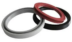
 Its unique design, which typically features a flexiblelip and a metal or plastic reinforcement ring, allows it to adapt to slight misalignments and shaft irregularities, enhancing its sealing effectiveness Its unique design, which typically features a flexiblelip and a metal or plastic reinforcement ring, allows it to adapt to slight misalignments and shaft irregularities, enhancing its sealing effectivenessoil seal 40 60 10.
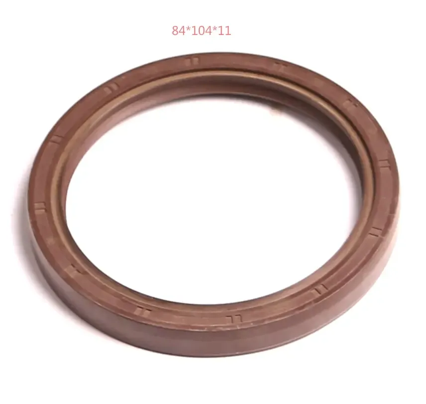
To prevent thelubricating oil from leaking outside even under high pressure of the oil.
Fluorine rubber (FKM, Viton™)
 The size of the gasket is typically specified by its inner diameter (ID), outer diameter (OD), and thickness The size of the gasket is typically specified by its inner diameter (ID), outer diameter (OD), and thicknessrectangular rubber gasket. The shape of the gasket can vary depending on the application, but common shapes include straight-sided, concave, convex, and angle-cut gaskets.
With spring Rubber O.D. wall Metal O.D. wall
The allowable total eccentricity is the maximum total eccentricity at which the sealing edge can accommodate shaft rotation and retain adequate sealing performance. The oil seal's allowable total eccentricity is affected by the design of the oil seal, the accuracy of the shaft, and the operating conditions.