The first study addressing the experimental convergence between in vitro spiking neurons and spiking memristors was attempted in 2013 (Gater et al., 2013). A few years later, Gupta et al. (2016) used TiO2 memristors to compress information on biological neural spikes recorded in real time. In these in vitro studies electrical communication with biological cells, as well as their incubation, was investigated using multielectrode arrays (MEAs). Alternatively, TiO2 thin films may serve as an interface material in various biohybrid devices. The bio- and neurocompatibility of a TiO2 film has been demonstrated in terms of its excellent adsorption of polylysine and primary neuronal cultures, high vitality, and electrophysiological activity (Roncador et al., 2017). Thus, TiO2 can be implemented as a nanobiointerface coating and integrated with memristive electronics either as a planar configuration of memristors and electrodes (Illarionov et al., 2019) or as a functionalization of MEAs to provide good cell adhesion and signal transmission. The known examples are electrolyte/TiO2/Si(p-type) capacitors (Schoen and Fromherz, 2008) or capacitive TiO2/Al electrodes (Serb et al., 2020). As a demonstration of the state of the art, an attempt at memristive interlinking between the brain and brain-inspired devices has been recently reported (Serb et al., 2020). The long-term potentiation and depression of TiO2-based memristive synapses have been demonstrated in relation to the neuronal firing rates of biologically active cells. Further advancement in this area is expected to result in scalable on-node processors for brain–chip interfaces (Gupta et al., 2016). As of 2017, the state of the art of, and perspectives on, coupling between the resistive switching devices and biological neurons have been reviewed (Chiolerio et al., 2017).


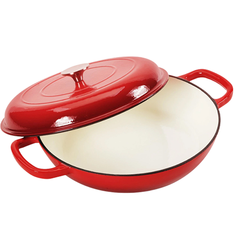
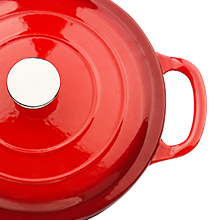
 From sautéing vegetables to searing steaks, the high walls of the skillet contain splatters and allow for a measure of depth that makes stirring and tossing a breeze From sautéing vegetables to searing steaks, the high walls of the skillet contain splatters and allow for a measure of depth that makes stirring and tossing a breeze
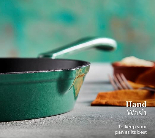
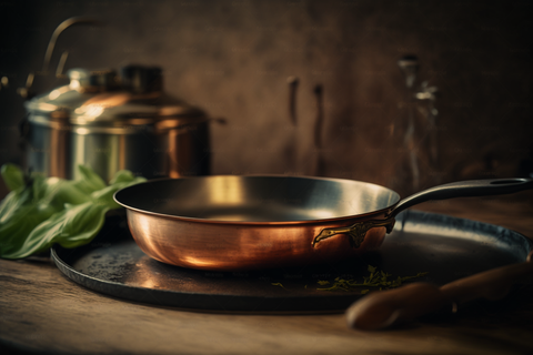 Copper core frying pans have a copper exterior for excellent heat conductivity and a stainless steel interior for durability and easy cleaning. They are ideal for cooking high-heat dishes, sauces, and eggs. However, they are expensive and prone to discolouration with prolonged use.